Cell Structure and Microscopy
Eukaryotic and Prokaryotic Cells
All living organisms are made of cells. There are two fundamental types of cell: eukaryotic cells and prokaryotic cells. Eukaryotic cells are found in animals and plants. They are complex, with a nucleus that holds the cell's genetic material. Prokaryotic cells, such as bacteria, are much smaller and simpler, with no true nucleus.
Animal cells contain the following sub-cellular structures:
- Nucleus — contains genetic material (DNA) that controls the cell's activities.
- Cell membrane — controls what enters and leaves the cell; it is selectively permeable.
- Mitochondria — the site of aerobic respiration, where energy is transferred from glucose.
- Ribosomes — tiny structures where proteins are synthesised (made).
Plant cells have all of these structures plus some additional ones:
- Cell wall — a rigid outer layer made of cellulose that provides support and protection.
- Chloroplasts — contain chlorophyll, the green pigment that absorbs light energy for photosynthesis.
- Vacuole — a large, permanent structure filled with cell sap that helps maintain the cell's shape.
Key Fact
Bacterial (prokaryotic) cells have chromosomal DNA that floats freely in the cytoplasm (no nucleus), plasmid DNA (small rings of extra DNA), a cell membrane, ribosomes, and some have a flagellum for movement. They do NOT have mitochondria or chloroplasts.
Specialised Cells
Cells become specialised to carry out particular functions. This process is called differentiation. The Edexcel specification requires you to know three examples of specialised cells.
Sperm cells are adapted for fertilisation:
- An acrosome at the tip releases enzymes to penetrate the egg cell.
- A haploid nucleus carries half the genetic information so a full set is restored at fertilisation.
- Many mitochondria in the middle section provide energy for the tail to swim.
- A long tail (flagellum) enables the sperm to move towards the egg.
Egg cells (ova) are adapted for fertilisation and early development:
- They contain nutrients in the cytoplasm to nourish the early embryo after fertilisation.
- They have a haploid nucleus, so the full diploid number is restored when the sperm fuses with the egg.
- After fertilisation, the cell membrane changes structure to prevent more than one sperm from entering. This is essential to maintain the correct chromosome number.
Ciliated epithelial cells line the airways (trachea and bronchi). They have tiny hair-like projections called cilia on their surface that beat rhythmically. This sweeps mucus (which has trapped dust and microorganisms) upwards towards the throat, keeping the lungs clean.
Key Fact
Cells become specialised through differentiation, so their structure is adapted to suit their function: sperm cells have a tail and many mitochondria for swimming, egg cells store nutrients for the embryo, and ciliated epithelial cells use cilia to sweep mucus out of the airways.
Microscopy
Cells are far too small to see with the naked eye, so scientists use microscopes to study them. A light microscope uses light and glass lenses. Light microscopes are cheap, portable and can show living cells in colour, but they only magnify up to about ×2000 and cannot show small sub-cellular structures clearly.
An electron microscope uses a beam of electrons instead of light. This gives a much higher magnification (over ×1,000,000) and a much higher resolution, so scientists can see tiny sub-cellular structures such as ribosomes and the internal detail of mitochondria. However, electron microscopes are large, very expensive, and the specimen must be dead.
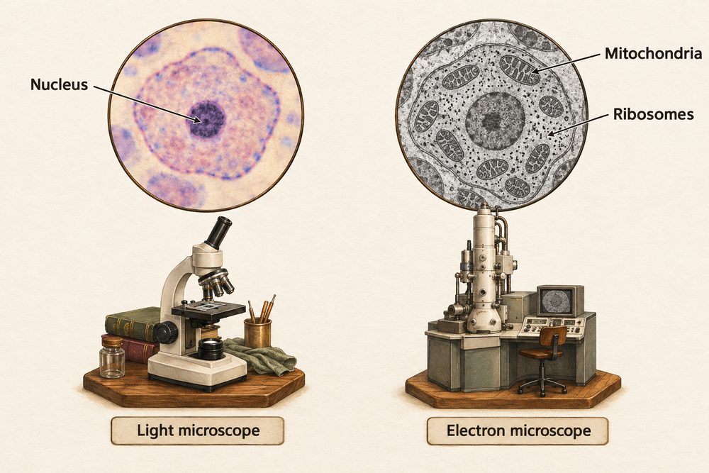
Resolution is the ability to tell apart two points that are very close together. Because electron microscopes have a much higher resolution than light microscopes, they reveal far more detail — this is what allowed scientists to discover sub-cellular structures.
Magnification tells you how many times larger an image is than the real object. It is calculated using the formula:
\(\text{magnification} = \frac{\text{image size}}{\text{real size}}\)
You can rearrange this to find the real size or the image size. Always make sure both measurements are in the same units before calculating — for example, convert millimetres (mm) to micrometres (µm) by multiplying by 1000.
For example, if a cell is 50 µm wide in real life and appears 100 mm wide in a photograph, first convert 100 mm to 100,000 µm, then magnification = 100,000 ÷ 50 = ×2000.
Key Fact
Magnification = image size ÷ real size. Electron microscopes have much higher magnification and resolution than light microscopes, so they can show sub-cellular structures. Always convert both measurements to the same units before you calculate.