Cell Structure and How We Know It
How We Came to Know the Cell
In 1665, Robert Hooke placed a thin slice of cork under a simple microscope and saw a pattern of tiny compartments. He called them “cells” because they reminded him of the small rooms in a monastery. Hooke could not see inside these compartments — his instrument was far too weak. Yet that single observation launched two centuries of investigation, ultimately revealing that cells are the fundamental unit of all life. The history of our understanding of cell structure is a story of technology driving science: each new generation of microscopes unlocked a new layer of biological reality.
This lesson explores two fundamental cell types — eukaryotic and prokaryotic — and traces how improvements in microscopy revealed the internal structures we now know so well.
Eukaryotic cells (animals, plants, fungi) have a true nucleus that encloses DNA within a nuclear membrane. Prokaryotic cells (bacteria) have no true nucleus — their DNA is a single loop in the cytoplasm. Prokaryotes are typically 10–100 times smaller than eukaryotes.
Organelles and Their Functions
Both animal and plant cells contain a core set of organelles whose design is closely matched to their function. The exam requires you to go beyond naming structures — you must explain why each structure’s shape, position or composition allows it to carry out its role efficiently.
Nucleus — contains the chromosomes (DNA) and controls the cell’s activities. The nuclear membrane keeps the genetic material separate from the cytoplasm. When a cell needs a particular protein, the genetic instructions are copied from DNA and sent out through pores in the nuclear membrane to the ribosomes.
Cell membrane — a thin, selectively permeable layer that controls which substances enter and leave. It contains receptor molecules that allow the cell to detect and respond to hormones and other chemical signals.
Mitochondria — the site of aerobic respiration. Their folded inner membranes (cristae) greatly increase the surface area available for respiration enzymes, making energy transfer more efficient. Cells with very high energy demands — such as muscle cells or sperm cells — are packed with mitochondria.
Ribosomes — tiny structures found in both the cytoplasm and on the endoplasmic reticulum. They are the site of protein synthesis. Ribosomes are present in both eukaryotic and prokaryotic cells, though prokaryotic ribosomes are smaller.
Plant cells have all the animal cell structures listed above, plus three additional ones:
Cell wall — made of cellulose, it surrounds the cell membrane and provides structural strength. It is fully permeable (allows all dissolved substances through) but prevents the cell from bursting when water enters by osmosis, keeping the cell turgid and supporting the plant.
Chloroplasts — contain the green pigment chlorophyll and are the site of photosynthesis. They are found mainly in cells near the surface of leaves and stems where light is available.
Permanent vacuole — a large fluid-filled space containing cell sap (dissolved sugars and salts). It presses the cytoplasm against the cell wall, keeping the cell turgid and helping to support the plant structure.
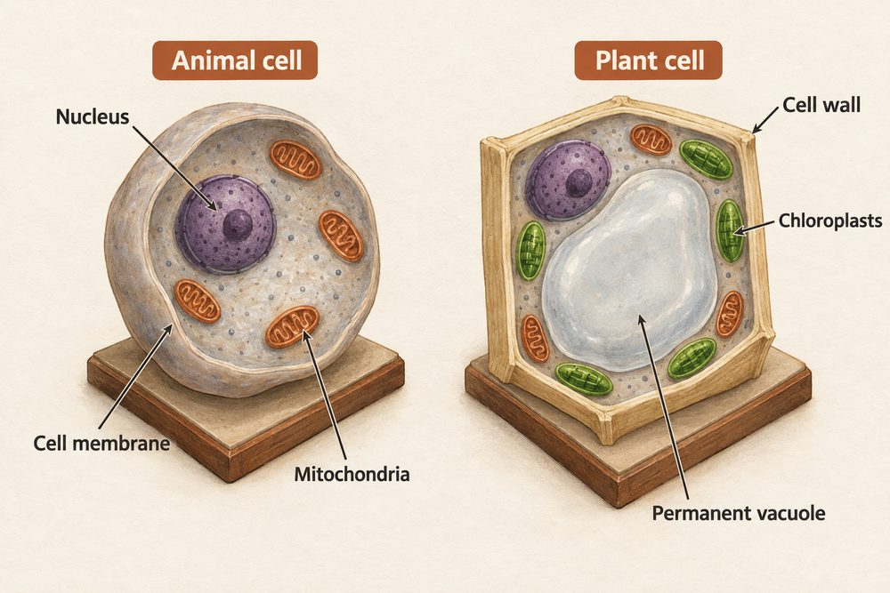
Plant cells have three additional structures compared to animal cells: cell wall (cellulose, structural support), chloroplasts (photosynthesis) and permanent vacuole (cell sap, turgidity). Bacterial cells have a cell wall but no true nucleus, no mitochondria and no chloroplasts.
Prokaryotic Cell Structure
Bacterial cells are prokaryotic: smaller, simpler and without membrane-bound organelles. Their genetic material is a single circular loop of DNA floating in the cytoplasm. Many bacteria also contain small, extra loops of DNA called plasmids, which can carry genes for antibiotic resistance and can be passed between bacteria. Bacteria do have ribosomes (for protein synthesis) and a cell wall (though made of a different material from plant cell walls, usually peptidoglycan).
From Hooke to the Electron Microscope
Light microscopes improved steadily after Hooke’s time. By the 19th century, biologists could see nuclei, cell divisions and the broad outlines of organelles. Light microscopes have a useful magnification of about ×1500 and a resolution of around 200 nm. To prepare a slide, a thin sample is placed on glass, stained (iodine to reveal plant cell walls and nuclei; methylene blue for animal cells), and covered with a coverslip lowered at an angle to avoid air bubbles.
The real breakthrough came in the 20th century with the invention of the electron microscope. Instead of light, it fires a beam of electrons at the specimen. This gives a resolution of around 0.2 nm — approximately 1,000 times better than a light microscope. Transmission electron microscopes (TEMs) can magnify specimens up to about two million times. This level of detail revealed the internal folds of mitochondria (cristae), the structure of ribosomes and the double membrane of the nucleus — all previously invisible. Each discovery confirmed and refined scientists’ models of cell structure.
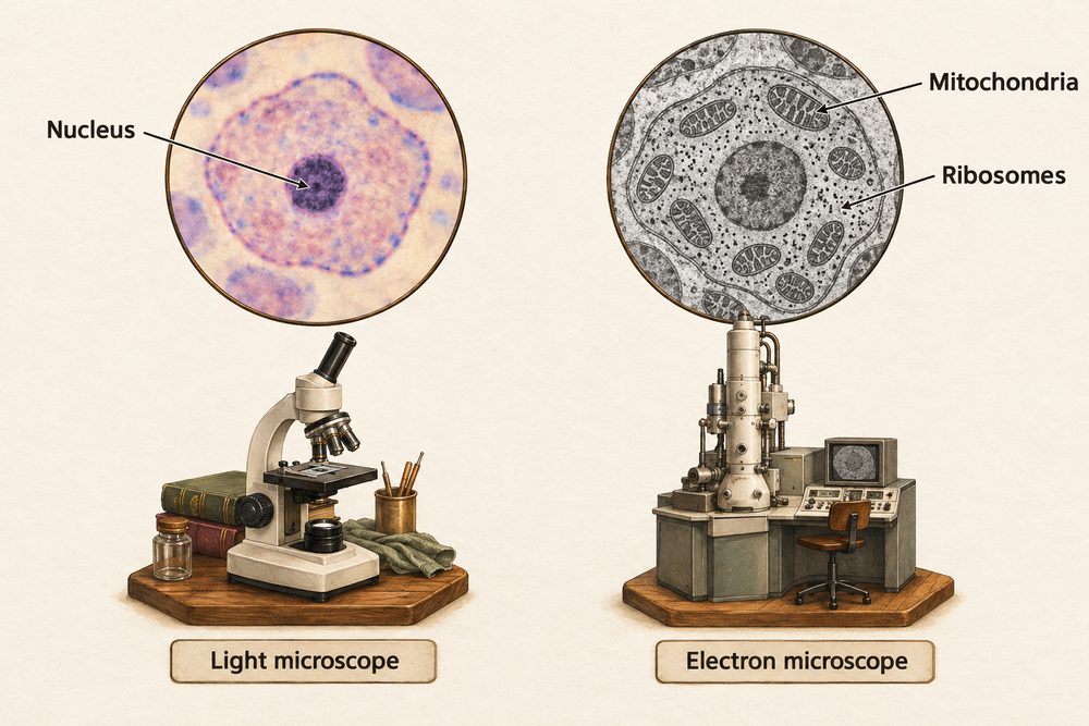
The development of electron microscopy is a clear example of how scientific knowledge develops with technology (a key Ideas about Science theme): new instruments allow observations that lead to revised or extended explanations. Hooke proposed cells existed; electron microscopists revealed how they worked from the inside.
The magnification formula is: $$\text{magnification} = \frac{\text{image size}}{\text{real size}}$$ Always convert both measurements to the same unit before dividing. Scientists often work in micrometres (µm) for cells and nanometres (nm) for organelles and molecules. \(1 \text{ mm} = 1000 \text{ µm} = 1{,}000{,}000 \text{ nm}\).
Worked example: A drawing of a cell measures 45 mm on the page. The actual cell is 0.015 mm. Magnification = \(\frac{45}{0.015} = \times 3000\). Always state the multiplication symbol and the number, e.g. ×3000. If asked to find the real size: real size = image size ÷ magnification.
Magnification = image size ÷ real size. Electron microscopes (~0.2 nm resolution, up to ×2,000,000) revealed mitochondrial cristae, ribosome structure and nuclear membrane detail that light microscopes (~200 nm resolution, ×1500) could not resolve.