Cell Structures and Microscopy
Eukaryotic and Prokaryotic Cells
All living organisms are made of cells. You need to understand two fundamental cell types: eukaryotic cells and prokaryotic cells.
Eukaryotic cells include animal and plant cells. They have a nucleus that encloses their genetic material within a nuclear membrane. They also contain membrane-bound organelles such as mitochondria and ribosomes. Prokaryotic cells — such as bacteria — are much smaller and simpler. They lack a true nucleus; instead, their genetic material is a single loop of DNA floating freely in the cytoplasm. Prokaryotes may also contain small rings of extra DNA called plasmids.
Eukaryotic cells (animal and plant) have a nucleus that encloses their DNA. Prokaryotic cells (bacteria) have no true nucleus — their DNA is a single loop in the cytoplasm, and they may also contain plasmids.
Sub-cellular Structures and Their Functions
Both animal and plant cells are eukaryotic, but they contain different sub-cellular structures. The exam requires you to explain how each structure's design relates to its function. This means you need to go beyond naming the structure — you must say why its shape, composition or position allows it to carry out its role efficiently.
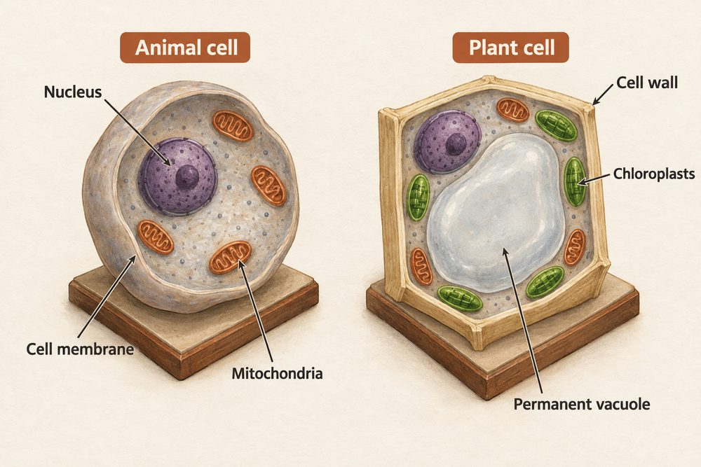
Nucleus — contains the cell's genetic material (chromosomes made of DNA) and controls the cell's activities by determining which proteins are made. The nuclear membrane keeps the DNA separate from the cytoplasm, protecting the genetic code.
Cytoplasm — a jelly-like substance where most chemical reactions take place. Enzymes in the cytoplasm control these metabolic reactions. It also contains structural proteins that give the cell its shape.
Cell membrane — a thin, selectively permeable layer that controls which substances enter and leave the cell. It contains receptor molecules that allow the cell to respond to chemical signals such as hormones and neurotransmitters.
Mitochondria — the site of aerobic respiration. They contain the enzymes needed for cellular respiration and transfer energy for the cell's processes. Cells with high energy demands (such as muscle cells) contain more mitochondria.
Ribosomes — tiny structures in the cytoplasm where protein synthesis occurs. They are found in both eukaryotic and prokaryotic cells, though they are smaller in prokaryotes.
Plant cells have all the structures listed above, plus three additional ones that reflect their roles in photosynthesis and structural support:
Chloroplasts — contain the green pigment chlorophyll and are the site of photosynthesis. They absorb light energy to convert carbon dioxide and water into glucose. Chloroplasts are found mainly in leaf cells and cells near the surface of stems, where light is available.
Permanent vacuole — a large, fluid-filled space containing cell sap (a solution of sugars and salts dissolved in water). It helps maintain the cell's rigidity by pressing the cytoplasm against the cell wall, keeping the cell turgid and supporting the plant structure.
Cell wall — made of cellulose and surrounds the cell membrane. It strengthens and supports the cell, preventing it from bursting when water enters by osmosis. Unlike the cell membrane, the cell wall is fully permeable and allows all dissolved substances through.
Plant cells have a cell wall (cellulose), chloroplasts (photosynthesis) and a permanent vacuole (cell sap) in addition to animal cell organelles. Bacterial cells have a cell wall, but no chloroplasts, no true nucleus and no mitochondria.
Microscopy
Microscopes allow us to see cells and sub-cellular structures invisible to the naked eye. You need to know about light microscopes and electron microscopes.
Light microscopes use glass lenses and visible light. They have a useful magnification of about \(\times 1500\) and a resolution of around 200 nm. To prepare a slide, place a thin sample on a glass slide, add a drop of stain (such as iodine or methylene blue) to make colourless structures visible, then lower a coverslip at an angle to avoid air bubbles.
You should be able to describe how to prepare slides, use staining techniques, and calculate magnification. Key steps:
1. Place a thin sample (e.g. onion epidermis or cheek cells) on a clean glass slide. 2. Add a drop of stain — iodine stains plant cell walls and nuclei yellow-brown, while methylene blue stains animal cell nuclei blue. 3. Gently lower a coverslip at 45° to avoid air bubbles. 4. Start on the lowest magnification objective lens, focus, then switch to higher magnifications.
To calculate the total magnification: \(\text{total magnification} = \text{eyepiece lens} \times \text{objective lens}\). For example, a \(\times 10\) eyepiece with a \(\times 40\) objective gives \(\times 400\) magnification.
Electron microscopes fire a beam of electrons at the specimen. A transmission electron microscope (TEM) has much higher resolution — down to about 0.2 nm — and a magnification of up to \(\times 2\,000\,000\). This allowed biologists to see sub-cellular structures like ribosomes and the internal folds of mitochondria (cristae) in fine detail, greatly increasing our understanding of how cells work.
The development of electron microscopy was a turning point in biology. Before electron microscopes, scientists could see cells and nuclei with light microscopes, but they could not resolve the detailed internal structure of organelles. Electron microscopy revealed that mitochondria have folded inner membranes (cristae) that increase the surface area for respiration enzymes, explaining why mitochondria are so efficient at energy transfer.
The magnification formula: \(\text{magnification} = \frac{\text{image size}}{\text{real size}}\). Always ensure both measurements are in the same units before calculating. Electron microscopes have much higher resolution (~0.2 nm) than light microscopes (~200 nm), allowing sub-cellular detail to be seen.