Cell Structure, Microscopy and Specialised Cells
Eukaryotic and Prokaryotic Cells
All living things are made of cells. Cells fall into two fundamental types: eukaryotic cells and prokaryotic cells. Knowing the differences between them — and being able to name sub-cellular structures — is tested across both papers of your Triple Biology exam.
Eukaryotic cells include animal and plant cells. They contain a nucleus that encloses their DNA, along with membrane-bound organelles such as mitochondria and ribosomes. Prokaryotic cells — bacteria — are much smaller and simpler. They have no true nucleus; instead, their genetic material is a single loop of DNA floating freely in the cytoplasm. Prokaryotes may also carry small rings of extra DNA called plasmids. Bacteria also have a cell wall, but it is made from a different material to plant cell walls.
Eukaryotic cells have a true nucleus, membrane-bound organelles and are typically 10–100 μm. Prokaryotic cells have no true nucleus, a single loop of DNA in the cytoplasm, and may contain plasmids. They are typically 1–5 μm.
Animal and Plant Cell Organelles
Both animal and plant cells are eukaryotic, but their organelle content differs. You must be able to name each organelle and link its structure to its function.
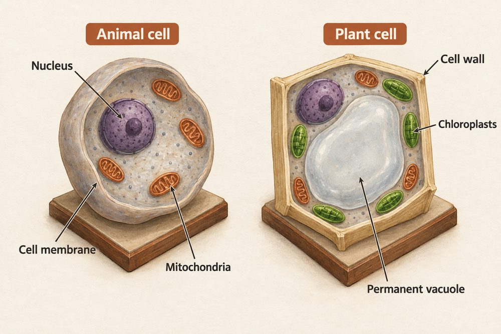
Nucleus — contains the cell’s DNA and controls the cell’s activities. It is enclosed by the nuclear envelope.
Cytoplasm — a jelly-like fluid where most chemical reactions take place, controlled by enzymes.
Cell membrane — a thin, selectively permeable layer that controls which substances enter and leave the cell.
Mitochondria — the site of aerobic respiration, where energy is transferred from glucose. Cells with high energy demands (e.g. muscle cells) contain more mitochondria.
Ribosomes — tiny structures where protein synthesis takes place. Found in both eukaryotic and prokaryotic cells.
Plant cells have all the organelles above, plus three additional structures:
Chloroplasts — contain the green pigment chlorophyll and are the site of photosynthesis.
Permanent vacuole — a large fluid-filled space containing cell sap (a dilute solution of sugars and salts). It keeps the cell rigid and supports the plant structure.
Cell wall — made of cellulose, it surrounds the cell membrane and provides structural strength. It prevents the cell from bursting when water enters by osmosis.
Plant cells have a cellulose cell wall, chloroplasts for photosynthesis and a permanent vacuole filled with cell sap. Animal cells have none of these. Both have a nucleus, mitochondria, ribosomes, cytoplasm and cell membrane.
Specialised Cells
Most cells are specialised — they have distinct structures matched to a specific job. Your exam frequently asks you to explain how a cell’s structure is adapted to its function.
Sperm cell — has a streamlined head with a pointed acrosome containing enzymes to digest the egg’s membrane. The midpiece is packed with mitochondria to supply energy for swimming. The long flagellum (tail) provides propulsion. The nucleus carries the male’s haploid genetic information to the egg.
Egg cell (ovum) — is the largest human cell and carries a haploid nucleus. It contains large food reserves (cytoplasm rich in nutrients) to nourish the developing embryo. Once fertilised, the cell membrane changes to prevent additional sperm from entering.
Ciliated epithelial cell — lines the trachea and bronchi. The surface is covered with tiny hair-like structures called cilia, which beat rhythmically to sweep mucus (containing trapped particles and bacteria) up towards the throat, keeping the airways clean.
Microscopy and Units
Microscopes let us see cells and sub-cellular structures too small for the naked eye. You must know two types: light microscopes and electron microscopes.
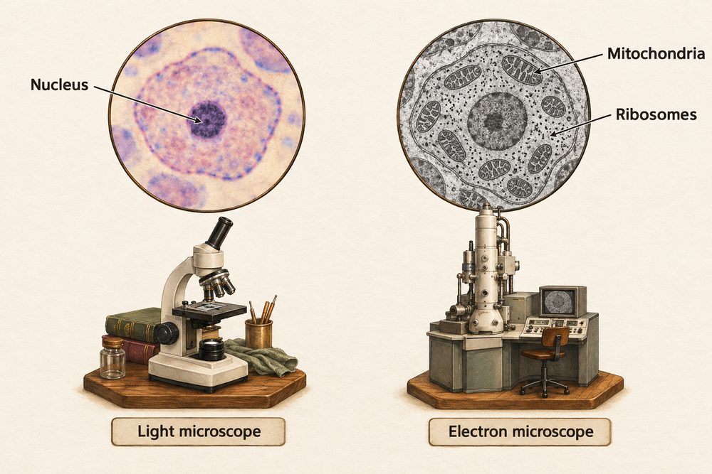
Light microscopes use visible light focused through glass lenses. They can magnify up to around ×1,500 with a resolution of about 200 nm. Electron microscopes fire a beam of electrons at the specimen; their resolution is about 0.2 nm and magnification can reach ×2,000,000. This revealed sub-cellular structures — such as ribosomes and the internal membranes of mitochondria — that light microscopes cannot resolve.
The magnification formula is:
$$\text{magnification} = \frac{\text{size of image}}{\text{size of real object}}$$
It can be rearranged to find real size or image size:
$$\text{real size} = \frac{\text{image size}}{\text{magnification}}$$
Example: A student draws a cell image that is 45 mm wide. The real cell is 0.045 mm (45 μm). What is the magnification?
$$\text{magnification} = \frac{45\,\text{mm}}{0.045\,\text{mm}} = \times 1{,}000$$
Always check both values are in the same units before dividing. Convert millimetres to micrometres if needed (1 mm = 1,000 μm).
Unit Conversions and Standard Form
Biology uses a range of units for measuring cells and structures. You must be confident with the following prefixes:
- milli (m) — one thousandth, \(10^{-3}\) of a metre. Typical scale for small organisms.
- micro (μ) — one millionth, \(10^{-6}\) of a metre. Typical for cells (10–100 μm).
- nano (n) — one billionth, \(10^{-9}\) of a metre. Used for sub-cellular structures and molecules.
- pico (p) — one trillionth, \(10^{-12}\) of a metre. Used for atoms and very small molecules.
Standard form expresses very large or very small numbers as \(A \times 10^n\). For example, a bacterial cell of \(2\,\mu\text{m}\) is \(2 \times 10^{-6}\,\text{m}\). Always express answers in standard form if the question uses it.
Key unit chain: 1 m = 1,000 mm = 1,000,000 μm = 1,000,000,000 nm. A typical animal cell is 10–30 μm; a bacterium is 1–5 μm; a ribosome is around 20 nm.