Nervous System, Reflexes and the Eye
Neurones and the Nervous System
The nervous system allows rapid communication across the body using electrical impulses. It has two main divisions: the central nervous system (CNS), consisting of the brain and spinal cord, and the peripheral nervous system, consisting of the nerves that connect the CNS to the body’s receptors and effectors.
There are three types of neurone:
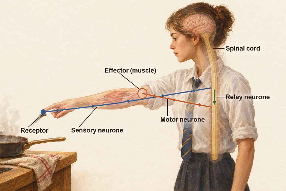
- Sensory neurone — carries impulses from receptors (e.g. in the skin, eyes, ears) towards the CNS. Has a long axon and a cell body located outside the spinal cord.
- Relay neurone — found entirely within the CNS. Connects sensory neurones to motor neurones and processes information within the spinal cord and brain.
- Motor neurone — carries impulses from the CNS to effectors (muscles or glands), causing a response.
Each neurone has a long axon surrounded by a myelin sheath in many neurones. The myelin sheath acts as an electrical insulator and dramatically increases the speed of impulse transmission. Damage to the myelin sheath (as in multiple sclerosis) slows impulse conduction.
Reflex arc sequence: receptor → sensory neurone → relay neurone (in spinal cord) → motor neurone → effector (muscle or gland). The myelin sheath insulates the axon and speeds up impulse transmission.
Synapses
Neurones do not physically touch each other. The tiny gap between two neurones is called a synapse. When an electrical impulse reaches the end of a neurone, it triggers the release of chemical messengers called neurotransmitters from small vesicles. These diffuse across the synaptic cleft and bind to complementary receptor proteins on the next neurone’s membrane. If enough neurotransmitter binds, a new electrical impulse is generated in the next neurone.
Synapses only allow impulses to travel in one direction (because neurotransmitters are released from one side only). Many drugs and toxins work by interfering with neurotransmitter release or reception at synapses.
A reflex is a fast, automatic response to a stimulus that does not require conscious thought. Reflexes bypass the brain, passing through the spinal cord instead, which is why they are so rapid.
Reflex arc pathway: stimulus (e.g. touching a hot surface) → receptor (pain receptors in the skin) → sensory neurone (impulse to spinal cord) → relay neurone (in the spinal cord) → motor neurone (impulse to effector) → effector (muscle contracts, hand withdrawn). The brain receives information about the reflex only after the response has already begun.
The Eye (Triple Only)
The eye is a sense organ that detects light. You must know the function of each structure and how the eye focuses light on the retina.
Cornea — the transparent curved cover at the front. It refracts (bends) most of the incoming light.
Iris — the coloured ring surrounding the pupil. Circular muscles (contract to constrict the pupil in bright light) and radial muscles (contract to dilate the pupil in dim light) control the amount of light entering.
Lens — a transparent flexible disc that fine-tunes the focus of light. It changes shape by the action of the ciliary muscles and suspensory ligaments.
Retina — the light-sensitive layer at the back of the eye. It contains rod cells (sensitive to dim light; detect black and white) and cone cells (sensitive to bright light; detect colour in three wavelengths: red, green, blue). Impulses travel along the optic nerve to the brain.
Accommodation
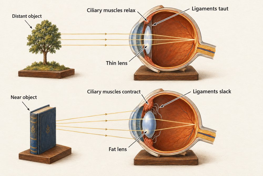
Accommodation is the eye’s ability to change focus between near and distant objects by altering the shape of the lens. When viewing a near object: the ciliary muscles contract, the suspensory ligaments loosen and the lens becomes rounder (more curved), increasing its refracting power. When viewing a distant object: the ciliary muscles relax, the suspensory ligaments pull taut and the lens flattens (less curved), reducing its refracting power.
Vision Defects and Correction
Short-sightedness (myopia) occurs when the eyeball is too long or the cornea is too curved. Light from distant objects is focused in front of the retina instead of on it. It is corrected with a concave (diverging) lens that spreads the light rays before they enter the eye.
Long-sightedness (hyperopia) occurs when the eyeball is too short or the cornea is too flat. Light from close objects is focused behind the retina. It is corrected with a convex (converging) lens that brings the light rays together before they enter the eye.
Cataracts form when the lens becomes cloudy, scattering light and reducing vision. The cloudy lens is surgically replaced with an artificial implant. Colour blindness results from a deficiency in one or more types of cone cell. It is sex-linked (carried on the X chromosome) and is much more common in males than females.
Short-sightedness: distant objects blurred; light focused in front of retina; corrected by concave lens. Long-sightedness: near objects blurred; light focused behind retina; corrected by convex lens.