Exchange, Transport and the Circulatory System
Surface Area to Volume Ratio
Single-celled organisms can exchange substances directly through their cell membrane because their surface area to volume ratio (SA:V ratio) is large. As organisms grow larger, their volume increases faster than their surface area, and the SA:V ratio falls. A small cube has a higher SA:V ratio than a large cube. For a large, active organism, the surface area of the body wall alone cannot exchange gases or nutrients fast enough — specialised exchange surfaces and transport systems are therefore essential.
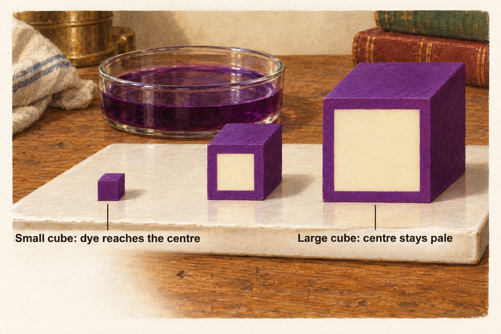
As organisms get larger, the surface area to volume ratio decreases. Large organisms need specialised exchange surfaces (e.g. alveoli, villi) and transport systems (e.g. blood) to supply all cells with oxygen and nutrients efficiently.
Alveoli and Gas Exchange
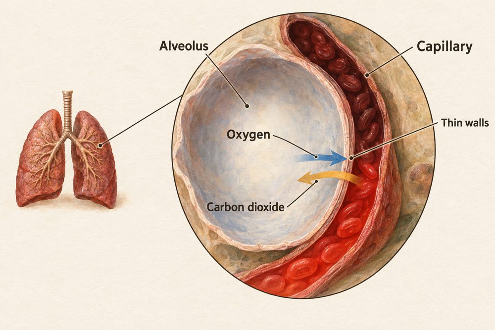
The lungs contain millions of tiny air sacs called alveoli (singular: alveolus). Their structure is adapted to maximise the rate of diffusion:
- Large surface area — millions of alveoli create an enormous total surface area for exchange.
- Thin walls — one cell thick, so the diffusion distance is very short.
- Good blood supply — a dense capillary network maintains a steep concentration gradient.
- Ventilation — breathing constantly refreshes air in the alveoli, maintaining a high oxygen concentration.
Fick’s law formalises the factors that affect diffusion rate: $$\text{Rate of diffusion} \propto \frac{\text{surface area} \times \text{concentration difference}}{\text{membrane thickness}}$$ This means the rate of diffusion increases when surface area is larger, concentration difference across the membrane is greater, or membrane thickness is smaller. The alveolus maximises all three: large surface area, steep gradient maintained by blood flow and ventilation, and walls only one cell thick.
Blood Components
Blood is a tissue transported around the body through blood vessels. It has four components:
- Red blood cells — contain haemoglobin, which binds to oxygen in the lungs (forming oxyhaemoglobin) and releases it in body tissues. Their biconcave disc shape increases surface area for gas exchange, and they have no nucleus to carry more haemoglobin.
- White blood cells — part of the immune system. Phagocytes engulf and digest pathogens; lymphocytes produce antibodies and provide immunological memory.
- Platelets — small cell fragments that trigger blood clotting at wound sites, forming a fibrin mesh that seals the injury and prevents blood loss and entry of pathogens.
- Plasma — the liquid part of blood, which transports dissolved substances including glucose, amino acids, carbon dioxide, urea and hormones.
Blood Vessels
There are three types of blood vessel, each adapted for its function:
- Arteries — carry blood away from the heart at high pressure. Their walls are thick and muscular with elastic fibres to withstand and smooth out the pressure pulses from the heartbeat.
- Veins — carry blood towards the heart at lower pressure. Their walls are thinner and they contain valves to prevent backflow.
- Capillaries — walls are only one cell thick, minimising diffusion distance. They form a dense network reaching every cell, allowing exchange of oxygen, glucose, carbon dioxide and waste with body tissues.
Arteries: high pressure, thick muscular walls, no valves, carry blood away from heart. Veins: low pressure, thin walls, valves prevent backflow, carry blood to heart. Capillaries: one-cell thick walls, site of exchange with tissues.
The Heart and Cardiac Output
The heart is a double pump. The right side pumps deoxygenated blood to the lungs; the left side pumps oxygenated blood to the rest of the body. It has four chambers: two upper atria that receive blood and two lower ventricles that pump it out. The left ventricle has a much thicker wall than the right because it must generate higher pressure to push blood around the entire body.
The output of the heart is described by the cardiac output equation: $$\text{cardiac output} = \text{stroke volume} \times \text{heart rate}$$ where cardiac output is measured in cm3 per minute, stroke volume is the volume pumped per beat (cm3) and heart rate is the number of beats per minute.
A student’s heart rate is 72 beats per minute and their stroke volume is 70 cm3. Calculate their cardiac output.
Cardiac output = stroke volume × heart rate = 70 × 72 = 5040 cm3 per minute (approximately 5 litres per minute). During vigorous exercise, both stroke volume and heart rate increase, so cardiac output can rise to over 20 litres per minute in a trained athlete.
Respiration
Aerobic respiration releases energy from glucose in the presence of oxygen: $$C_6H_{12}O_6 + 6O_2 \rightarrow 6CO_2 + 6H_2O$$ This occurs in the mitochondria and is the main source of energy for all cell processes.
When oxygen is limited (e.g. during intense exercise), cells switch to anaerobic respiration. In human muscle cells, glucose is partially broken down to produce lactate (also called lactic acid). This releases much less energy than aerobic respiration and causes muscle fatigue. After exercise, extra oxygen is needed to break down accumulated lactate — this is the oxygen debt.
In yeast and plant cells, anaerobic respiration produces ethanol and carbon dioxide — a process called fermentation. Fermentation is used commercially in baking (CO2 makes bread rise) and in brewing (ethanol is produced).