Nervous System and Reflexes
Structure of the Nervous System
The human nervous system allows the body to detect changes in the environment and produce coordinated responses. It has two main divisions:
- Central nervous system (CNS) — the brain and spinal cord. This is the processing centre: it receives information from receptors, interprets it and sends out instructions to effectors.
- Peripheral nervous system — the nerves that extend from the CNS to the rest of the body. These include sensory neurones (carrying signals to the CNS) and motor neurones (carrying signals from the CNS to muscles and glands).
The nervous response pathway: stimulus → receptor → sensory neurone → relay neurone (CNS) → motor neurone → effector → response. The CNS (brain and spinal cord) is the coordination centre in the middle of every voluntary response.
Neurones and Their Structure
There are three types of neurone:
- Sensory neurones carry impulses from receptors to the CNS.
- Relay neurones are found within the CNS and connect sensory neurones to motor neurones.
- Motor neurones carry impulses from the CNS to effectors (muscles, which contract, or glands, which secrete).
All neurones have a cell body (containing the nucleus), dendrites (short extensions that receive impulses) and a long axon that transmits impulses away from the cell body. Many axons are surrounded by a myelin sheath — an insulating fatty layer that speeds up the transmission of electrical impulses.
Neurones do not physically touch each other. The tiny gap between two neurones is called a synapse. When an electrical impulse reaches the end of one neurone, it triggers the release of chemical messengers called neurotransmitters into the synaptic gap. These molecules diffuse across the gap and bind to receptor proteins on the next neurone, triggering a new electrical impulse there.
Synapses are important because they allow signals to pass in only one direction (neurotransmitters are released from one side only), and because they allow the nervous system to integrate information from many inputs before deciding whether to generate a response. Many drugs and toxins work by interfering with synaptic transmission — for example, some drugs mimic neurotransmitters, while others block their receptors.
Reflex Actions
A reflex action is an automatic, rapid response to a stimulus that bypasses conscious thought. Reflexes protect the body from harm — for example, pulling your hand away from a hot surface, blinking when something approaches your eye, or the pupil reflex in bright light.
The pathway for a reflex is called the reflex arc. In a spinal reflex, the relay neurone is in the spinal cord rather than the brain, which is why reflexes are so fast — the signal does not need to travel all the way to the brain for a decision to be made.
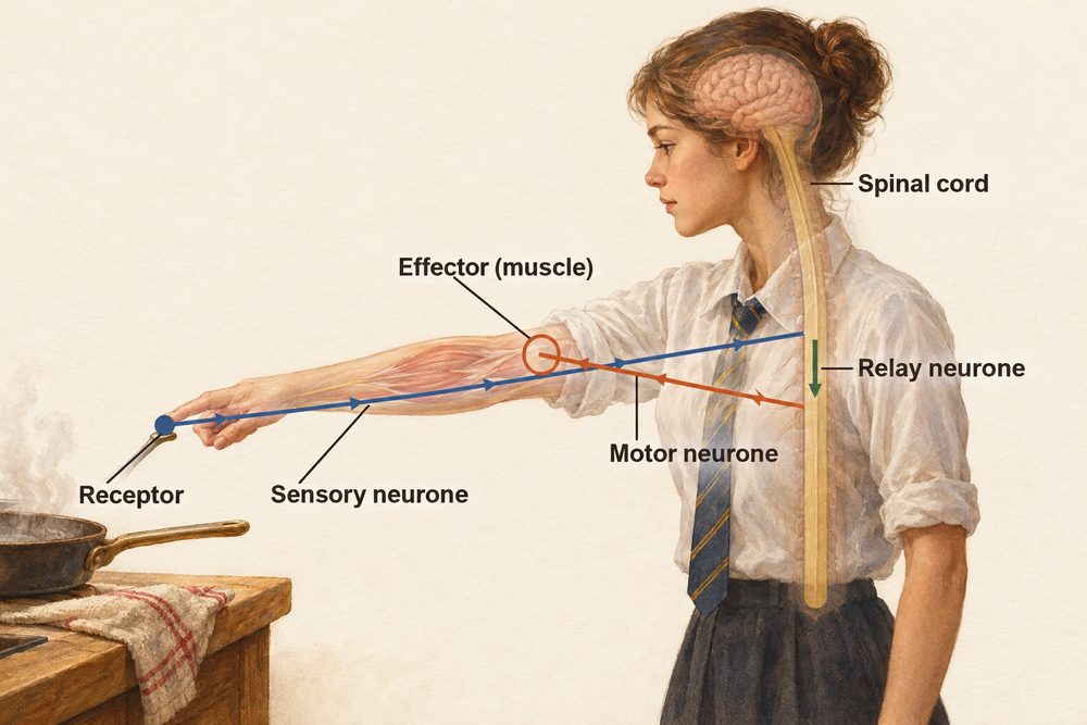
Reflex arcs bypass the brain — the relay neurone is in the spinal cord — making reflexes faster than voluntary actions. This speed is crucial: reflexes protect us from harm before we have time to think. Examples include withdrawing from pain and the pupil light reflex.
The Reflex Arc: Step by Step
Using the example of pulling your hand away from a hot surface:
- A receptor in the skin (a thermoreceptor) detects the intense heat stimulus.
- An impulse travels along a sensory neurone to the spinal cord.
- In the spinal cord, the impulse crosses a synapse and passes along a relay neurone.
- The relay neurone crosses another synapse and triggers a motor neurone.
- The impulse reaches the effector (the bicep muscle in the arm), which contracts.
- The response: the hand is pulled away. Simultaneously, a signal is sent to the brain so you become conscious of the event — but this happens after the response has already occurred.
When bright light shines into the eye, the pupil automatically constricts (gets smaller) to protect the sensitive retina. This is the pupil light reflex. Light-sensitive receptors in the retina send signals along sensory neurones to the brain stem; motor signals return along motor neurones to the circular muscles of the iris, causing them to contract and the pupil to narrow. In dim light, the opposite occurs: radial muscles contract and the pupil dilates to allow more light in.
The pupil reflex is coordinated in the brain stem rather than the spinal cord — it is still a reflex (automatic) but its relay station is higher up the nervous system. Doctors use this reflex to assess brain function: an absence of the pupil reflex after a head injury may indicate serious brain damage.
The Brain: Regions and Functions
The brain is the master coordination centre of the nervous system. It is made up of billions of interconnected neurones and is divided into distinct regions, each associated with particular functions. The cerebral cortex is the outer layer of the cerebrum, the largest part of the brain. It is responsible for intelligence, memory, language and consciousness. The cerebellum coordinates balance and the precision of movement. The brain stem (which includes the medulla) connects the brain to the spinal cord. It controls automatic functions such as heart rate and breathing rate.
Understanding the brain remains one of the biggest challenges in science. Techniques such as functional MRI (fMRI) scanning allow scientists to observe which brain regions are active during different tasks, but the complexity of billions of neurones interacting makes a full understanding difficult. Investigations into brain function are constrained by ethical limits on what experiments can be performed on living human subjects, by individual variation between brains, and by the fact that damage to one brain region may produce different effects in different people. This is an ongoing area of scientific investigation where models are continually refined as new evidence emerges.