Plant and Animal Transport Systems
Why Do Large Organisms Need Transport Systems?
Single-celled organisms can rely on diffusion alone to supply all their needs — their high surface area to volume (SA:V) ratio means diffusion distances are short. But as organisms get larger, their SA:V ratio decreases, and diffusion becomes too slow to supply oxygen and nutrients to cells deep inside the body, or to remove waste products efficiently. Multicellular organisms therefore evolved dedicated transport systems alongside specialised exchange surfaces.
Plant Transport — Xylem and Phloem
Plants have two types of transport tissue bundled together in vascular bundles: xylem and phloem.
Xylem vessels transport water and dissolved minerals upwards from roots to leaves. The cells are dead and form hollow tubes with thick walls reinforced with lignin, which makes them rigid and waterproof and provides structural support for the plant. Water moves through xylem as an unbroken column pulled upwards by evaporation from the leaves — this is the transpiration stream.
Phloem tubes transport dissolved sugars (mainly sucrose) from the leaves — where they are made by photosynthesis — to the rest of the plant, including roots, growing tips and storage organs. This process is called translocation. Unlike xylem, phloem cells are living and have sieve plates (pores) to allow the flow of sucrose. Translocation requires energy and can move substances both up and down the plant.
Xylem: dead hollow tubes reinforced with lignin; transports water and minerals upwards. Phloem: living tubes with sieve plates; transports dissolved sugars (sucrose) up and down by translocation. Both found in vascular bundles.
Transpiration
Transpiration is the evaporation of water vapour from the leaves through tiny pores called stomata. As water evaporates from the leaf surface, more water is drawn up through the xylem from the roots — creating the transpiration stream. Guard cells open and close the stomata: they become turgid (absorb water, swell, open stoma) in light, and flaccid (lose water, close stoma) in darkness or drought conditions.
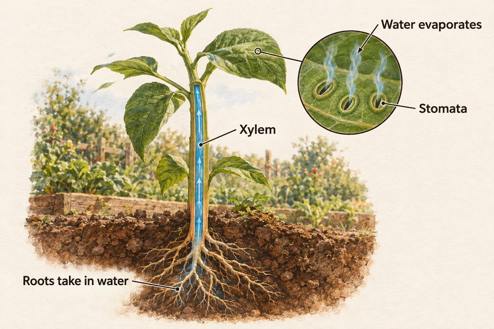
Four environmental factors increase the rate of transpiration:
Light intensity — causes stomata to open wider, increasing water loss. Temperature — increases the kinetic energy of water molecules, speeding up evaporation. Wind/air movement — removes humid air from around the leaf, maintaining a steep concentration gradient for water vapour to diffuse out. Humidity — low humidity (dry air) increases the concentration gradient, speeding up water loss; high humidity slows it.
A potometer is used to measure the rate of water uptake by a plant cutting, which is taken as a proxy for the rate of transpiration. The distance moved by an air bubble in a capillary tube per unit time is recorded as conditions are varied.
The Human Circulatory System
Humans have a double circulatory system: one circuit pumps blood from the heart to the lungs for gas exchange (pulmonary circuit), and the other pumps oxygenated blood from the heart to the body (systemic circuit). This maintains high blood pressure for efficient delivery of oxygen and nutrients.
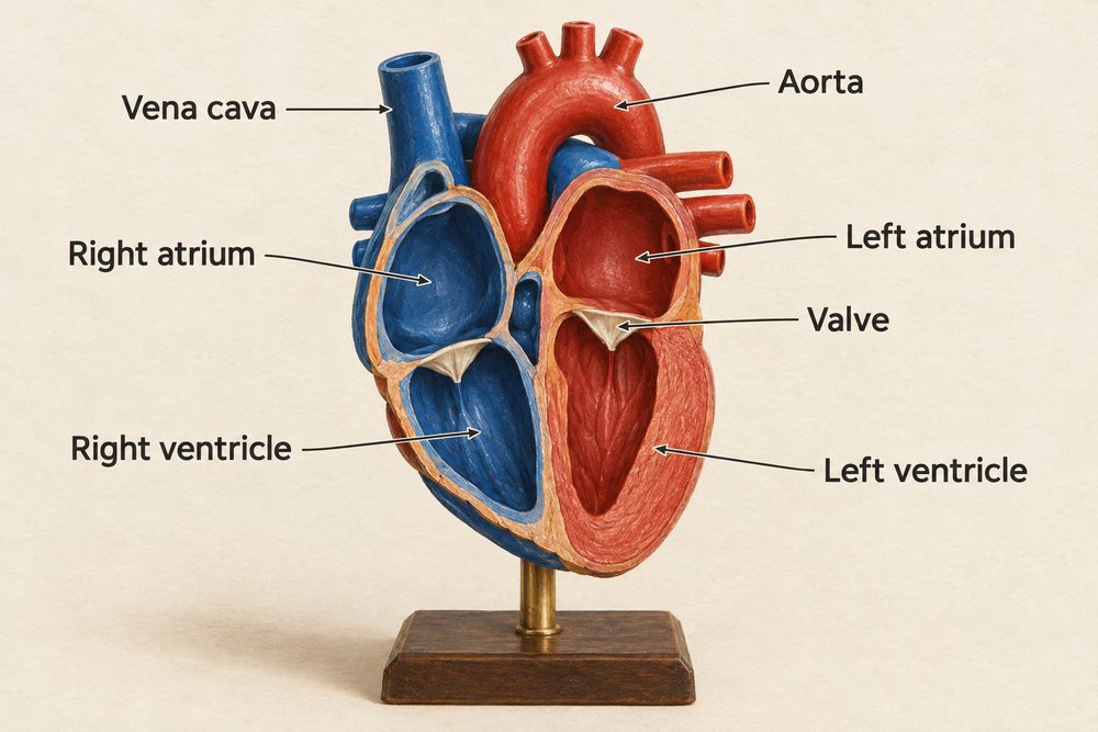
The heart has four chambers — two atria (receive blood) and two ventricles (pump blood out). The right side pumps deoxygenated blood to the lungs; the left side pumps oxygenated blood to the body. The left ventricle has a thicker muscular wall because it needs to generate higher pressure to pump blood around the whole body. Valves prevent backflow and ensure blood flows in one direction.
Arteries carry blood away from the heart at high pressure. They have thick, muscular walls with elastic fibres to withstand the surge of blood with each heartbeat. Their lumen (internal channel) is relatively small.
Veins carry blood back to the heart at low pressure. They have thinner walls and a larger lumen. They contain valves to prevent backflow and ensure blood moves only towards the heart.
Capillaries are microscopic vessels with walls just one cell thick, allowing efficient exchange of substances by diffusion between the blood and surrounding tissues. Oxygen and glucose diffuse out; carbon dioxide and waste diffuse in.
Red blood cells are biconcave discs with no nucleus, packed with haemoglobin to carry oxygen. Their biconcave shape increases surface area for oxygen absorption. White blood cells are part of the immune system — phagocytes engulf pathogens; lymphocytes produce antibodies. Platelets are cell fragments that help blood to clot at wound sites. Plasma is the liquid component of blood that transports dissolved substances including glucose, amino acids, urea, hormones, carbon dioxide and antibodies.
Blood flow through the double circulatory system: vena cava → right atrium → right ventricle → pulmonary artery → lungs → pulmonary vein → left atrium → left ventricle → aorta → body. The left ventricle has thicker walls than the right because it pumps blood further.
Gas Exchange — The Lungs
The lungs are the site of gas exchange between the blood and the air. They contain millions of tiny air sacs called alveoli. Alveoli are adapted for efficient gas exchange with features that are the same as other exchange surfaces: very large total surface area, walls just one cell thick to minimise diffusion distance, moist inner surface to allow gases to dissolve, and a rich network of capillaries to maintain steep concentration gradients by removing oxygen and delivering carbon dioxide.
The Digestive System
The digestive system breaks down large, insoluble food molecules into small, soluble ones that can be absorbed into the bloodstream. Key organs include the stomach (acid environment, protease enzymes break down proteins), the small intestine (enzymes from the pancreas complete digestion; products absorbed through villi into the blood), and the large intestine (water reabsorbed). The liver produces bile, which emulsifies fats (breaks large fat droplets into smaller ones, increasing the surface area for lipase enzymes to act on).
The inner wall of the small intestine is lined with millions of finger-like projections called villi, each covered in even smaller microvilli (the brush border). Together these massively increase the surface area for absorption. Each villus contains a dense capillary network (for absorbing glucose and amino acids into the blood) and a lacteal (a lymph vessel for absorbing fatty acids and glycerol from digested fats).
Villi share the same adaptations as other exchange surfaces: large surface area, thin walls (one cell thick), and a good blood supply to maintain steep concentration gradients. Glucose and amino acids are absorbed by a combination of diffusion and active transport; water is absorbed by osmosis.