The Nervous System and Reflex Arc
Structure of the Nervous System
The human nervous system allows the body to detect changes in the environment (stimuli) and produce rapid, coordinated responses. It consists of the central nervous system (CNS) — the brain and spinal cord — and the peripheral nervous system — nerves that connect the CNS to receptors and effectors throughout the body (B3.1a).
The pathway for any nervous response follows a clear sequence: stimulus → sensory receptor → sensory neurone → CNS (relay neurone) → motor neurone → effector → response.
The nervous pathway: stimulus → receptor → sensory neurone → relay neurone (in CNS) → motor neurone → effector → response. The CNS (brain and spinal cord) processes information and coordinates the response.
Neurones and Their Structure
There are three types of neurone. Sensory neurones carry impulses from receptors to the CNS. Relay neurones are found within the CNS and link sensory neurones to motor neurones. Motor neurones carry impulses from the CNS to effectors (muscles or glands).
Both sensory and motor neurones share common structural features: a cell body containing the nucleus, short dendrites that receive incoming impulses, and a long axon that carries the electrical impulse away from the cell body. In motor neurones the long axon runs directly from the spinal cord to the effector. In sensory neurones a long dendron connects the receptor to the cell body, with a shorter axon entering the CNS. The axon is often surrounded by a myelin sheath, which insulates the axon and greatly increases the speed of impulse transmission.
Synapses
Where two neurones meet there is a tiny gap called a synapse. An electrical impulse cannot cross this gap directly. Instead, when an impulse arrives at the end of the pre-synaptic neurone, neurotransmitters are released from vesicles into the synaptic cleft. These chemicals diffuse across the gap and bind to specific receptors on the post-synaptic neurone, triggering a new electrical impulse that continues the pathway.
Synapses ensure impulses travel in one direction only, because neurotransmitters are released from one side and receptors are only present on the other. They also allow one neurone to connect with several others simultaneously, enabling complex coordination throughout the nervous system (B3.1b).
A reflex arc is the pathway that produces a rapid, automatic, protective response without conscious thought (B3.1c). The pathway goes: receptor → sensory neurone → relay neurone (in spinal cord) → motor neurone → effector. Crucially, the signal passes through the spinal cord rather than the brain, making the response faster than a voluntary reaction.
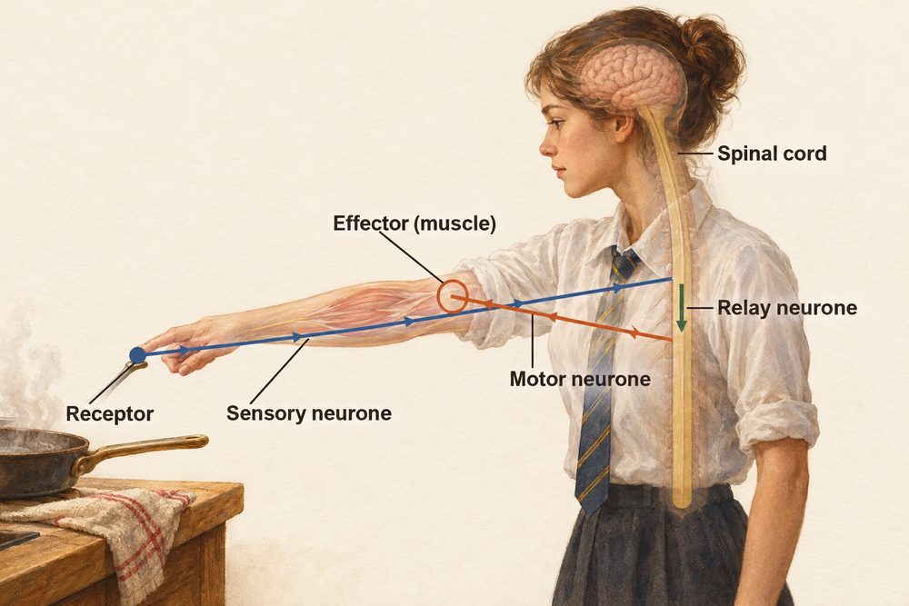
For example, when your fingertip touches something sharp: pain receptors detect the stimulus → a sensory neurone carries the impulse to the spinal cord → a relay neurone connects directly to a motor neurone → the motor neurone stimulates arm muscles to contract → the hand is withdrawn. This protects the body from harm before conscious awareness of pain. Other reflex examples include the pupil reflex (pupils constrict in bright light) and the knee-jerk reflex.
Reflex arcs produce rapid, automatic responses that bypass the brain and pass through the spinal cord instead. This makes reflexes faster than voluntary responses and protects the body from injury before conscious thought.
The ruler drop test is a simple practical used to measure voluntary reaction time (B3.1c practical context, PAG B6). One person holds a ruler vertically at the 0 cm mark; the subject places their thumb and finger at the base without touching it. When the ruler is released without warning, the subject catches it as quickly as possible. The shorter the distance fallen, the faster the reaction time.
To improve reliability: repeat at least five times, discard anomalous results, and calculate a mean. Variables that could affect results include caffeine, fatigue, distractions and practice. This experiment measures a voluntary response rather than a true reflex — the information passes through the brain (visual cortex → motor cortex) rather than being processed in the spinal cord.
The Brain
The brain is the main coordination centre of the nervous system. It contains billions of neurones connected by synapses. Five parts are named in the specification (B3.1f): the cerebrum, the cerebellum, the medulla, the hypothalamus and the pituitary gland.
Cerebral cortex — the highly folded outer layer of the cerebrum, responsible for consciousness, intelligence, memory, language and personality. The folding greatly increases surface area, allowing more neurones to be packed in. Different regions of the cortex are specialised — for example, the visual cortex processes information from the eyes, and the motor cortex controls voluntary muscle movements.
Cerebellum — located at the back of the brain. It coordinates balance, fine motor control and smooth, precise movements. Damage to the cerebellum results in loss of coordination and unsteady movement.
Medulla oblongata — at the base of the brain where it connects to the spinal cord. It controls involuntary, life-sustaining processes such as breathing rate, heart rate and swallowing. Because these functions are automatic, the medulla keeps them running without conscious thought. The hypothalamus sits above the medulla. It monitors body temperature and water balance, and it controls the pituitary gland just below it. The pituitary gland releases hormones that control other endocrine glands, which is why it is called the master gland.
Our understanding of brain function comes from several sources: studying patients with brain injuries, using electroencephalography (EEG) to record electrical activity, and modern MRI scanning to map active brain regions. However, the brain is enormously complex — many functions involve multiple regions working together, and some functions remain poorly understood. This makes treating brain disorders challenging.
The Eye
The eye is a complex sensory receptor for light (B3.1d). The key structures to know are: the cornea (refracts light), the iris (controls pupil size and therefore light entering), the lens (focuses light onto the retina), and the retina (converts light into electrical impulses via rod and cone photoreceptors). Impulses travel to the brain via the optic nerve.
Accommodation is the ability of the eye to focus on objects at different distances by changing the shape of the lens. To focus on a near object: the ciliary muscles contract, the suspensory ligaments slacken, and the lens becomes fatter (more curved) to refract light more strongly. To focus on a distant object: the ciliary muscles relax, the suspensory ligaments pull taut, and the lens becomes thinner (less curved), refracting light less.
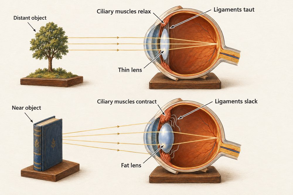
Myopia (short-sightedness): distant objects appear blurred because the eyeball is too long or the lens too curved, causing light to focus in front of the retina. Corrected with a concave (diverging) lens. Hyperopia (long-sightedness): near objects are blurred because the eyeball is too short or lens too flat, causing light to focus behind the retina. Corrected with a convex (converging) lens. Both can now also be corrected with laser eye surgery, which reshapes the cornea.