Cell Structure and Microscopy
Eukaryotic and Prokaryotic Cells
All living things are made of cells. Cells are divided into two fundamental types: eukaryotic cells and prokaryotic cells. Understanding the differences between these two types is essential biology knowledge and is regularly tested in exams.
Eukaryotic cells include both animal and plant cells. They have a nucleus that encloses their genetic material (DNA), along with other membrane-bound organelles such as mitochondria and ribosomes. Prokaryotic cells — bacteria — are much smaller and simpler. They lack a true nucleus: instead, their genetic material is a single loop of DNA that floats freely in the cytoplasm. Prokaryotes may also contain small rings of extra DNA called plasmids.
Eukaryotic cells (animal and plant) have a nucleus that encloses their DNA. Prokaryotic cells (bacteria) have no true nucleus — their DNA is a single loop in the cytoplasm, and they may also contain plasmids.
A typical animal cell is around 10–30 µm (micrometres) in diameter. A prokaryotic bacterial cell is much smaller, typically 1–5 µm. You need to be comfortable converting between units and using standard form. The key prefixes are: centi (\(10^{-2}\)), milli (\(10^{-3}\)), micro (\(10^{-6}\)) and nano (\(10^{-9}\)).
Animal and Plant Cells
Both animal and plant cells are eukaryotic, but they contain different organelles. You must be able to name each organelle and explain how its structure relates to its function.
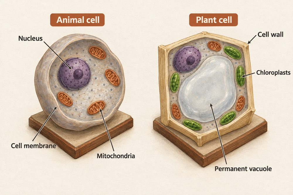
Nucleus — contains the cell’s genetic material (DNA) and controls the cell’s activities. The DNA carries the instructions for making proteins.
Cytoplasm — a jelly-like substance where most chemical reactions take place. Enzymes in the cytoplasm control these metabolic reactions.
Cell membrane — a thin layer that surrounds the cell and controls which substances enter and leave. It is selectively permeable.
Mitochondria — the site of aerobic respiration, where energy is transferred from glucose. Cells that need lots of energy (such as muscle cells) contain more mitochondria.
Ribosomes — tiny structures in the cytoplasm where protein synthesis occurs. They are found in both eukaryotic and prokaryotic cells.
Plant cells have all of the organelles listed above, plus three additional structures:
Chloroplasts — contain the green pigment chlorophyll and are the site of photosynthesis. They absorb light energy to make glucose.
Permanent vacuole — a large, fluid-filled space containing cell sap (a weak solution of sugars and salts). It helps keep the cell rigid and supports the plant.
Cell wall — made of cellulose and surrounds the cell membrane. It strengthens and supports the cell, preventing it from bursting when water enters by osmosis.
Plant cells have a cell wall (cellulose), chloroplasts (photosynthesis) and a permanent vacuole (cell sap) in addition to the organelles found in animal cells. Bacterial cells have a cell wall but no chloroplasts, no true nucleus and no mitochondria.
Cell Specialisation
Not every cell in an organism looks the same. Cells become specialised — they develop particular structures that allow them to perform a specific function. The exam regularly asks you to explain how a cell’s structure relates to its function.
Sperm cell — has a streamlined head, a long tail (flagellum) for swimming, and many mitochondria to provide energy for movement. It carries the male DNA to the egg.
Nerve cell (neurone) — has a long axon to carry electrical impulses over long distances, branched endings (dendrites) to connect with other nerve cells, and a myelin sheath to insulate the signal and speed up transmission.
Muscle cell — contains many mitochondria to supply energy for contraction. Muscle cells are elongated so they can contract and relax to produce movement.
Red blood cell — has a biconcave disc shape to increase surface area for absorbing oxygen. It has no nucleus, leaving more room to carry haemoglobin.
Root hair cell — has a long, thin extension (the root hair) that increases the surface area for absorbing water and mineral ions from the soil by osmosis and active transport.
Xylem cell — a hollow, dead tube reinforced with lignin that transports water and mineral ions from roots to leaves in a continuous column. The hollow centre (lumen) allows uninterrupted flow.
Phloem cell — has sieve plates with tiny pores that allow dissolved sugars to flow through the plant from leaves to other parts. This movement is called translocation.
Cell Differentiation
The process by which a cell becomes specialised is called differentiation. As a cell differentiates, it acquires the sub-cellular structures it needs for its function.
Most animal cells differentiate at an early stage of development — during embryonic growth. Once they have specialised, they generally cannot change into a different cell type, and cell division in mature animals is mainly for repair and replacement. Plant cells, however, retain the ability to differentiate throughout their lives. Undifferentiated plant cells are found in regions called meristems, located at the tips of roots and shoots. This is why you can take a cutting from a plant and it will grow into a new individual.
Most animal cells differentiate early in development and are then permanently specialised. Plant cells can differentiate throughout life because they retain meristem tissue — undifferentiated cells at root and shoot tips.
Microscopy
Microscopes allow us to see cells and sub-cellular structures that are invisible to the naked eye. There are two main types you need to know: light microscopes and electron microscopes.
Light microscopes use glass lenses and visible light to produce a magnified image. They have a maximum useful magnification of about \(\times 1{,}500\) and a resolution of around 200 nm. Light microscopes were first developed in the 1590s and allowed scientists to discover cells for the first time.
Electron microscopes fire a beam of electrons at the specimen rather than using light. They have a much higher resolution (down to about 0.2 nm) and a useful magnification of up to \(\times 2{,}000{,}000\). This allows biologists to see sub-cellular structures such as ribosomes and the internal structure of mitochondria in much finer detail. The development of electron microscopes in the 1930s was a turning point — it revealed structures inside cells that no one had seen before.
Light microscopes: max magnification \(\approx \times 1{,}500\), resolution \(\approx 200\) nm. Electron microscopes: max magnification \(\approx \times 2{,}000{,}000\), resolution \(\approx 0.2\) nm. Higher resolution means finer detail can be distinguished.
You must be able to use and rearrange the magnification formula:
$$\text{magnification} = \frac{\text{size of image}}{\text{size of real object}}$$
This can be rearranged to find the real size or the image size:
$$\text{real size} = \frac{\text{image size}}{\text{magnification}}$$
$$\text{image size} = \text{magnification} \times \text{real size}$$
Worked example: A student measures the image of a cell as 30 mm across. The real cell is 0.03 mm (30 µm). Calculate the magnification.
$$\text{magnification} = \frac{30}{0.03} = \times 1{,}000$$
Always check both measurements are in the same units before dividing. Express very large or very small answers in standard form where appropriate.
Using a Light Microscope
You need to be able to use a light microscope to observe, draw and label plant and animal cells, and calculate magnification from a drawing. Here is the standard procedure:
Step 1: Place a thin specimen (e.g. a layer of onion epidermis or a cheek cell smear) on a clean glass slide.
Step 2: Add a drop of water (for plant cells) or a stain such as iodine (to make structures more visible). Methylene blue is used for animal cells.
Step 3: Lower a coverslip at an angle using a mounted needle to avoid trapping air bubbles, which distort the image.
Step 4: Clip the slide onto the microscope stage and select the lowest power objective lens first.
Step 5: Use the coarse focus knob to bring the image roughly into focus, then the fine focus knob to sharpen it.
Step 6: To see more detail, switch to a higher power objective lens and refocus using the fine adjustment only.
Drawing rules: Use a sharp pencil. Draw clear, continuous lines (no shading). Label key structures with straight ruled lines. Include a title, the magnification used, and a scale bar.
When using a light microscope: always start on the lowest power objective, use a stain to make structures visible, lower the coverslip at an angle to avoid air bubbles, and include a magnification scale on your biological drawing.
When answering questions on cell structure, always link the organelle to its function — for example, “mitochondria are the site of aerobic respiration, which transfers energy for the cell.” In magnification calculations, the most common mistake is forgetting to convert units so that both image size and real size are in the same unit (usually mm or µm) before dividing. Always show every step of your working — you can pick up method marks even if the final answer is wrong. For Required Practical 1, be ready to describe the method and explain why each step matters (e.g. “the coverslip is lowered at an angle to prevent air bubbles, which would distort the image”).