Culturing Microorganisms and Plant Disease
Culturing Microorganisms
Microorganisms such as bacteria can be grown in the laboratory using nutrient media. This allows scientists to study how they grow, test antibiotics, and investigate disease.
An aseptic technique is used to prevent contamination of bacterial cultures by unwanted microorganisms from the environment. This ensures that only the microorganism being studied grows on the culture medium.
To culture bacteria, scientists use either a nutrient agar plate or a nutrient broth. Agar plates provide a solid surface on which individual colonies can be seen, while broth allows bacteria to grow in liquid suspension.
Aseptic Technique Steps
To prevent contamination, a specific set of precautions must be followed during any work with microorganisms:
- Sterilise all equipment (Petri dishes, inoculating loops) before use — an inoculating loop is passed through a Bunsen burner flame until it glows red.
- Lids of Petri dishes are secured with tape but not sealed completely, to allow oxygen in but prevent contamination from above.
- Work near a Bunsen flame to create an updraught that carries airborne microorganisms away from the open plates.
- Incubate cultures at a maximum of 25 °C in schools (to reduce the risk of growing pathogens harmful to humans). In industry, cultures can be incubated at higher temperatures, often around 37 °C.
In separate GCSE Biology you may be asked to describe how to investigate the effect of antiseptics or antibiotics on bacterial growth. Paper discs soaked in different concentrations of antiseptic are placed on an inoculated agar plate. After incubation, clear zones called zones of inhibition show where bacteria could not grow.
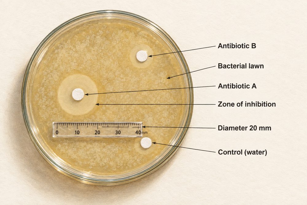
The area of the zone of inhibition can be calculated using \(A = \pi r^2\), where \(r\) is the radius of the clear zone. A larger zone means the antiseptic is more effective at killing or preventing bacterial growth.
Plant Disease
Plants can be infected by a range of pathogens including viruses, bacteria, fungi, and protists. These diseases reduce crop yields and can devastate agriculture.
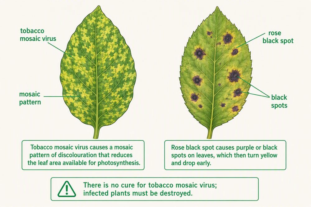
Tobacco mosaic virus (TMV) causes a mosaic pattern on leaves, reducing the area available for photosynthesis. There is no cure — infected plants must be destroyed.
Examples of Plant Diseases
Rose black spot is a fungal disease that causes purple or black spots on leaves. Affected leaves turn yellow and drop early, reducing the plant’s ability to photosynthesise. It spreads through water and wind and can be treated with fungicides or by removing and destroying affected leaves.
Crown gall disease is caused by the bacterium Agrobacterium tumefaciens, which inserts its DNA into the plant cells and causes tumour-like growths (galls) on stems and roots.
Plant diseases can be identified by:
- Stunted growth — nutrient deficiency or pathogen infection.
- Spots on leaves — fungal or bacterial infection (e.g. rose black spot).
- Areas of decay or rot — fungal or bacterial infection.
- Discolouration — viral infection (e.g. TMV mosaic pattern) or mineral deficiency.
- Malformed stems or leaves — viral infection or pest damage.
Gardeners and farmers can also use testing kits, laboratory analysis, or reference material to confirm diagnoses.
Plant Defence Responses
Plants have evolved physical, chemical, and mechanical defences against pathogens and herbivores:
- Physical barriers: Cellulose cell walls, tough waxy cuticles on leaves, and bark on trees prevent pathogen entry.
- Chemical defences: Some plants produce antimicrobial substances (e.g. mint and witch hazel) or poisons to deter herbivores.
- Mechanical adaptations: Thorns, hairs, and leaves that droop or curl when touched deter herbivores.
Plants cannot move away from threats, so their defences must be structural or chemical. Some plants seal off infected areas by producing callose deposits to block the spread of pathogens through the phloem.